Growth Scan and Doppler in Pregnancy: Fetal Growth, Blood Flow & Fetal Growth Restriction Explained
A growth scan checks how your baby is growing, while Doppler ultrasound looks at blood flow between the placenta and baby. Here is what these scans can tell you and what happens when a baby measures small.
Pregnancy scans do more than provide a picture of your baby. At different stages of pregnancy, ultrasound can answer very different questions.
A growth scan is mainly used to assess how your baby is growing. It looks at measurements such as the baby's head, abdomen and thigh bone and uses these measurements to estimate fetal weight.
A Doppler ultrasound looks at blood flow. Depending on the clinical situation, it can help doctors assess circulation in vessels such as the umbilical artery and understand how the placenta and baby are functioning.
These two assessments are often discussed together, particularly when there is concern that a baby may be smaller than expected or that the placenta may not be working as well as it should.
But there is an important point that parents sometimes miss:
A baby measuring small does not automatically mean that something is wrong.
Some babies are simply constitutionally small. Others may have fetal growth restriction (FGR), where the baby has not reached its expected growth potential because of an underlying problem.
Understanding that difference is one of the main reasons growth is followed over time.
What is a growth scan in pregnancy?
A growth scan is an ultrasound examination used to assess fetal growth and development later in pregnancy.
During the scan, the sonologist or fetal medicine specialist takes several measurements of the baby. These commonly include:
Head circumference
Biparietal diameter
Abdominal circumference
Femur length
Estimated fetal weight
The measurements are compared with expected values for the baby's gestational age.
Importantly, the estimated weight shown on the scan is not the baby's actual weight. It is a calculated estimate based on ultrasound measurements.
Fetal growth assessment is also more meaningful when measurements are considered over time. A single scan provides a snapshot; sequential assessments can show whether the baby is following an expected growth pattern.
When is a growth scan recommended?
Not every pregnant woman needs the same number of growth scans.
A growth scan may be recommended when your obstetrician or fetal medicine specialist wants to check fetal growth more closely. This can happen for several reasons, including:
The baby appears smaller or larger than expected
There is a previous history of fetal growth restriction
Maternal medical conditions may affect placental function
There are concerns about the placenta
There are concerns about amniotic fluid
Blood pressure or other pregnancy complications develop
Previous ultrasound findings require follow-up
The pregnancy is considered higher risk
There are twins or multiple fetuses
Earlier measurements suggest that additional monitoring may be useful
The timing and frequency depend on the individual pregnancy. There is no single schedule that applies to everyone.
What does a growth scan measure?
The scan does not simply ask, “How much does the baby weigh?”
It looks at several measurements because fetal growth is a pattern rather than one number.
Head measurements
Measurements of the baby's head help assess growth of the skull and brain.
Abdominal circumference
The abdominal circumference is particularly useful when assessing fetal size and growth. Changes in abdominal growth can sometimes provide important clues about fetal growth restriction.
Femur length
The length of the thigh bone is another measurement used in fetal biometry.
Estimated fetal weight
The measurements are combined using an ultrasound formula to calculate an estimated fetal weight (EFW).
The result is then compared with reference values for the baby's gestational age.
What does a fetal growth percentile mean?
You may see a percentile written on your ultrasound report, such as the 10th, 25th, 50th or 75th percentile.
A percentile compares the baby's estimated size with babies of the same gestational age in the reference population used by the reporting system.
For example, a baby around the 50th percentile is close to the middle of that reference range.
A lower percentile does not automatically mean that the baby is unhealthy.
This is where the distinction between small for gestational age (SGA) and fetal growth restriction (FGR) becomes important.
What is the difference between a small baby and fetal growth restriction?
A baby may be considered small for gestational age when measurements such as estimated fetal weight or abdominal circumference fall below a particular percentile, commonly the 10th percentile.
But SGA and FGR are not interchangeable terms.
Some babies are naturally small and remain healthy. In other pregnancies, a smaller size reflects a problem with the baby's growth potential, often related to placental function.
Fetal growth restriction refers to a situation in which the fetus has not achieved its expected growth potential because of a pathological process.
That distinction is why doctors do not diagnose FGR simply by looking at one number. Growth pattern, Doppler findings and the wider clinical picture matter.
Why does a baby become growth restricted?
There is no single cause of fetal growth restriction.
One important factor is placental insufficiency, where the placenta is not delivering oxygen and nutrients as effectively as expected.
Other factors can include:
Maternal medical conditions
Hypertensive disorders of pregnancy
Certain placental problems
Fetal chromosomal or structural abnormalities
Certain infections
Multiple pregnancy
Other maternal, fetal or placental factors
Sometimes, despite appropriate investigation, a single clear cause is not identified.
This is one reason fetal growth monitoring needs to be individualized rather than based only on the estimated fetal weight.
What is a Doppler scan in pregnancy?
A Doppler ultrasound is a specialized form of ultrasound used to assess blood flow.
Instead of only producing an anatomical image, Doppler evaluates movement of blood through vessels.
Different Doppler techniques can provide information about blood flow, including spectral Doppler and colour flow imaging.
In pregnancy, Doppler assessment can be particularly useful when there is a concern about fetal growth or placental function.
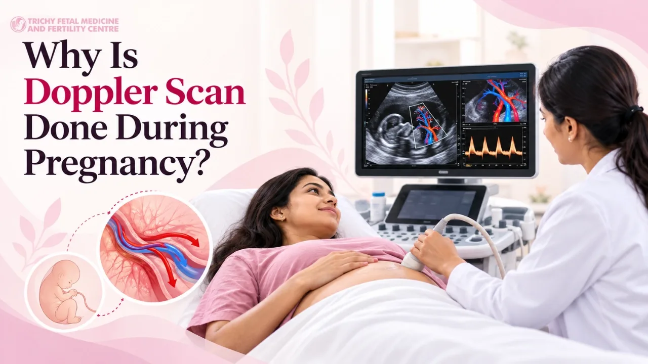
Why is Doppler done during pregnancy?
Doppler can provide information about the circulation between the mother, placenta and baby.
When a baby is suspected to have fetal growth restriction, the umbilical artery Doppler is particularly important in many clinical protocols.
It can help doctors assess resistance to blood flow through the placenta and determine how closely the pregnancy may need to be monitored.
In pregnancies complicated by FGR, guidelines recommend serial Doppler assessment of the umbilical artery as part of fetal surveillance, with the frequency depending on the severity and Doppler findings.
Doppler is therefore not simply another measurement of the baby's size. It answers a different question.
Growth scan: How is the baby growing?
Doppler: What does the blood flow tell us about the circulation supporting the baby?
Which blood vessels can be assessed with Doppler?
The vessel examined depends on the clinical question.
Umbilical artery
The umbilical artery carries blood from the fetus toward the placenta.
Umbilical artery Doppler is particularly important when fetal growth restriction is suspected because it can provide information about placental resistance.
Middle cerebral artery
The middle cerebral artery is a blood vessel in the baby's brain.
In selected situations, its blood-flow pattern may provide additional information about fetal circulation.
Uterine arteries
Uterine artery Doppler assesses blood flow in the maternal vessels supplying the uterus and placenta.
It may be useful in particular pregnancy assessments, depending on the clinical context.
Ductus venosus
The ductus venosus is a fetal blood vessel that connects with the fetal circulation near the heart.
Assessment of this vessel is relevant in selected high-risk situations, although Doppler protocols vary according to the clinical indication and guideline being followed.
Not every pregnancy requires assessment of all these vessels.
Does an abnormal Doppler mean that the baby is in danger?
Not necessarily.
A Doppler result needs to be interpreted according to which vessel was examined, the gestational age, the specific Doppler measurement and the rest of the pregnancy assessment.
An abnormal result may mean that closer monitoring is needed. It does not, by itself, tell the entire story.
This is particularly important when parents see a result on a report and try to interpret it without the surrounding clinical information.
Your doctor may compare:
Current growth measurements
Previous growth scans
Doppler findings
Amniotic fluid
Fetal movements
Maternal blood pressure
Gestational age
Other pregnancy-specific findings
The next step is based on the complete picture.
How is Doppler related to fetal growth restriction?
There is a close relationship between fetal growth and placental blood flow.
When the placenta is not functioning normally, the baby may receive less oxygen and nutrients than expected. The baby may then show slower growth.
Doppler can sometimes identify changes in circulation associated with placental dysfunction.
This makes Doppler particularly useful when FGR has been diagnosed or is strongly suspected. However, Doppler is not a standalone test for diagnosing FGR.
A baby can be small with a reassuring Doppler, and Doppler findings may change the way a pregnancy with growth restriction is monitored.
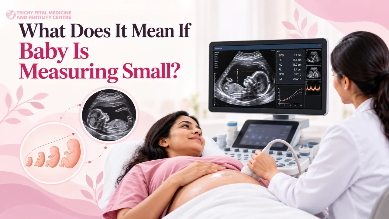
What does it mean if the baby is measuring small?
Hearing that your baby is “small” can understandably cause concern.
But a small measurement is not automatically a diagnosis of fetal growth restriction.
Your doctor may first look at:
How small the baby is.
Whether the abdominal circumference or estimated weight is below a relevant threshold.
Whether the baby has followed a similar growth pattern on previous scans.
Whether Doppler blood flow is reassuring.
Whether amniotic fluid is normal.
Whether there are maternal health issues.
Whether there are any other ultrasound findings.
Sometimes the appropriate next step is simply to repeat the growth assessment after an interval.
In other pregnancies, additional surveillance may be needed.
Why are repeat growth scans sometimes necessary?
Fetal growth is dynamic.
A single ultrasound cannot tell your doctor exactly how quickly your baby is growing.
If a baby measures smaller than expected, a repeat scan can show whether growth continues along a similar trajectory or whether there has been a change.
This is particularly useful because growth velocity can provide information that a single measurement cannot.
The interval between scans matters too. Performing growth scans too close together may not provide a meaningful indication of growth because ultrasound measurements themselves have a margin of error.
Your doctor therefore chooses the timing based on the pregnancy and the reason for monitoring.
Can a growth scan predict my baby's exact birth weight?
No.
A growth scan provides an estimated fetal weight, not an exact measurement.
Ultrasound-based fetal weight calculations have inherent limitations, and the estimated weight can differ from the baby's actual birth weight.
For this reason, doctors usually use the estimated weight as one piece of clinical information rather than treating it as an exact prediction.
A scan saying that a baby weighs a particular amount does not mean the baby will be born at precisely that weight.
Growth scan vs Doppler: what is the difference?
Growth scan vs Doppler: what is the difference?
Growth Scan
Doppler Scan
Assesses fetal size and growth
Assesses blood flow
Measures fetal body parts
Measures blood-flow characteristics
Calculates estimated fetal weight
Provides information about circulation
Helps assess growth pattern
Helps assess placental/fetal circulation
Useful when growth is a concern
Particularly useful in selected high-risk pregnancies
Can be repeated to monitor growth
May be repeated to monitor changes in blood flow
Growth scan vs Doppler: what is the difference?
Growth Scan: Assesses fetal size and growth
Doppler Scan: Assesses blood flow
Growth Scan: Measures fetal body parts
Doppler Scan: Measures blood-flow characteristics
Growth Scan: Calculates estimated fetal weight
Doppler Scan: Provides information about circulation
Doppler Scan: Particularly useful in selected high-risk pregnancies
Growth Scan: Can be repeated to monitor growth
Doppler Scan: May be repeated to monitor changes in blood flow
Growth scan vs Doppler: what is the difference?
The two tests can complement one another, but they are not interchangeable.
Can Doppler alone diagnose fetal growth restriction?
No.
Fetal growth restriction is assessed using fetal biometry and the overall clinical picture. Doppler can provide important additional information, especially about placental and fetal circulation.
In other words, Doppler helps answer:
“How is the circulation supporting this baby?”
The growth scan helps answer:
“How is this baby growing?”
Putting those findings together gives the doctor a much clearer picture than either measurement alone.
What happens if fetal growth restriction is confirmed?
The next step depends on several factors.
There is no single treatment plan for every pregnancy with FGR.
Monitoring may include:
Repeat growth assessment
Umbilical artery Doppler
Other fetal surveillance when indicated
Assessment of amniotic fluid
Maternal blood pressure monitoring
Assessment of fetal movements
Additional specialist review
Planning the timing and mode of delivery when appropriate
The aim is to balance the risks of continuing the pregnancy against the risks associated with delivering too early.
The exact approach depends on gestational age, fetal growth, Doppler findings, fetal wellbeing and maternal condition. Professional guidelines differ in some details, so management is individualized.
Does fetal growth restriction always mean there is a serious problem?
No.
The severity and significance of FGR vary from one pregnancy to another.
Some babies with restricted growth remain stable with monitoring for a considerable period. In other pregnancies, worsening Doppler findings or other concerns may require more intensive surveillance or earlier delivery.
This is why doctors do not make decisions based solely on the baby's estimated weight.
A small baby with reassuring findings may be managed very differently from a baby with evidence of significant placental dysfunction.
Can a small baby catch up in growth?
Some babies who measure small during pregnancy are constitutionally small and continue to grow normally.
Others may have genuine growth restriction and require closer surveillance.
Whether a baby continues to grow appropriately depends on the underlying reason for the small measurements and how the pregnancy progresses.
That is why trend matters.
A sequence of appropriately timed growth scans can be more informative than one isolated measurement.
What factors can affect fetal growth?
Fetal growth is influenced by many factors, including:
Genetic factors
Placental function
Maternal health
Maternal blood pressure
Certain pregnancy complications
Multiple pregnancy
Fetal conditions
Environmental and other factors
Sometimes a baby's size simply reflects familial or constitutional characteristics.
A smaller baby therefore should not automatically be assumed to have a disease.
What is early-onset and late-onset fetal growth restriction?
Fetal growth restriction can present at different stages of pregnancy.
Early-onset FGR develops earlier in pregnancy and may be associated with more significant placental or fetal problems.
Late-onset FGR occurs later in pregnancy and can be more subtle. A baby may not appear dramatically small, which is one reason clinical assessment and appropriate surveillance remain important.
The distinction is based on gestational age and clinical criteria, rather than simply whether the baby appears small on ultrasound.
Does every small baby need a Doppler scan?
Not necessarily.
The need for Doppler depends on why the baby is measuring small and what other findings are present.
Your doctor may consider:
The estimated fetal weight
Abdominal circumference
Growth trajectory
Gestational age
Previous scan results
Maternal conditions
Placental findings
Other fetal assessments
If there is a concern about fetal growth restriction, Doppler may become an important part of monitoring.
The appropriate test should be based on the clinical situation rather than performed simply because the baby is below an arbitrary number.
How often do you need a growth scan in a high-risk pregnancy?
There is no universal schedule.
A woman with a pregnancy that is considered high risk may require additional scans, but the timing depends on the reason for the increased risk.
If FGR has been diagnosed, professional guidance commonly recommends serial monitoring, including umbilical artery Doppler at intervals determined by the clinical findings.
Your obstetrician or fetal medicine specialist can determine the appropriate schedule for your pregnancy.
What happens during a growth scan with Doppler?
The examination is performed using ultrasound.
You will generally lie on an examination couch while the ultrasound probe is moved over your abdomen with ultrasound gel.
The clinician takes the necessary fetal measurements and may assess:
Baby's head
Abdomen
Femur
Estimated fetal weight
Amniotic fluid
Placenta
Fetal position
Relevant blood vessels using Doppler
The exact components depend on why the scan has been requested.
You may hear Doppler sounds during the examination. These sounds represent blood-flow information and are not the baby's heartbeat being “recorded” in the way a conventional audio recording works.
Is a growth scan with Doppler safe?
Ultrasound does not use ionizing radiation.
Diagnostic ultrasound is widely used during pregnancy when medically indicated. Doppler is also an ultrasound-based technique.
As with any medical examination, ultrasound should be used appropriately and for a clinical purpose, with the examination performed by trained professionals.
Can a growth scan detect every pregnancy problem?
No.
A growth scan is designed primarily to assess fetal growth and related findings. It is not a substitute for every other pregnancy screening or diagnostic test.
Depending on the stage of pregnancy and the clinical situation, different examinations may be used to assess:
Fetal anatomy
Chromosomal risk
Fetal heart structure
Brain and nervous system
Placental function
Fetal growth
Blood flow
Maternal health
A normal growth scan therefore does not mean that every possible fetal or maternal condition has been excluded.
When should you discuss a growth scan with a fetal medicine specialist?
A fetal medicine consultation may be considered when there is a specific concern about fetal growth, Doppler findings or another pregnancy-related issue.
Examples include:
The baby is measuring significantly smaller than expected
Serial scans show reduced growth
Doppler findings are abnormal
There is maternal hypertension or another medical condition affecting pregnancy
There is a history of FGR
There are concerns about the placenta
A twin pregnancy requires specialist monitoring
Another ultrasound finding requires expert assessment
A specialist can review the scan alongside previous measurements and the clinical history rather than interpreting one number on its own.
What should you ask your doctor after a growth scan?
If you have been told that your baby is small or that Doppler monitoring is needed, it can help to ask specific questions.
For example:
What is my baby's estimated weight?
What percentile is the baby measuring at?
Has the growth changed compared with the previous scan?
Is the abdominal circumference within the expected range?
Is the Doppler normal?
Which blood vessel was assessed?
Does the baby meet criteria for fetal growth restriction?
When should the next scan be performed?
Do I need additional fetal monitoring?
Are there any maternal conditions affecting the baby's growth?
At what point would delivery need to be considered?
Having these answers can make the report much easier to understand.
Growth monitoring is about more than one number
One of the most important things to remember is that fetal growth is a process, not a single measurement.
A baby who measures on the smaller side may be perfectly healthy.
Another baby with a similar estimated weight may need closer monitoring because the growth pattern, Doppler findings or other clinical features are different.
That is why fetal medicine specialists look at the whole picture.
Growth measurements tell us about size. Doppler adds information about circulation. Serial assessments show how things are changing.
Together, these findings can help guide appropriate monitoring during pregnancy.
Being told that your baby is “small” can be unsettling, especially when the ultrasound report contains numbers and terms that are difficult to interpret.
Try not to judge the result from the estimated weight or percentile alone.
A fetal growth assessment is most useful when the measurements are interpreted in context. Your doctor may look at previous scans, growth trends, Doppler blood flow, amniotic fluid, maternal health and other findings before deciding whether further monitoring is needed.
If your pregnancy requires a growth scan or Doppler assessment, the purpose is not simply to produce another set of numbers. These examinations help your care team understand how your baby is growing and, when necessary, whether the circulation supporting the baby needs closer observation.
For patients seeking fetal medicine consultation and pregnancy scan services in Trichy, Trichy Fetal Medicine and Fertility Centre provides services including growth scans with Doppler, fetal echocardiography, anomaly scans, NIPT, genetic counselling and other fetal medicine services.
To arrange this at our Trichy centre, see growth scan with Doppler; if you are unsure whether you need it, our doctors can tell you after a review.