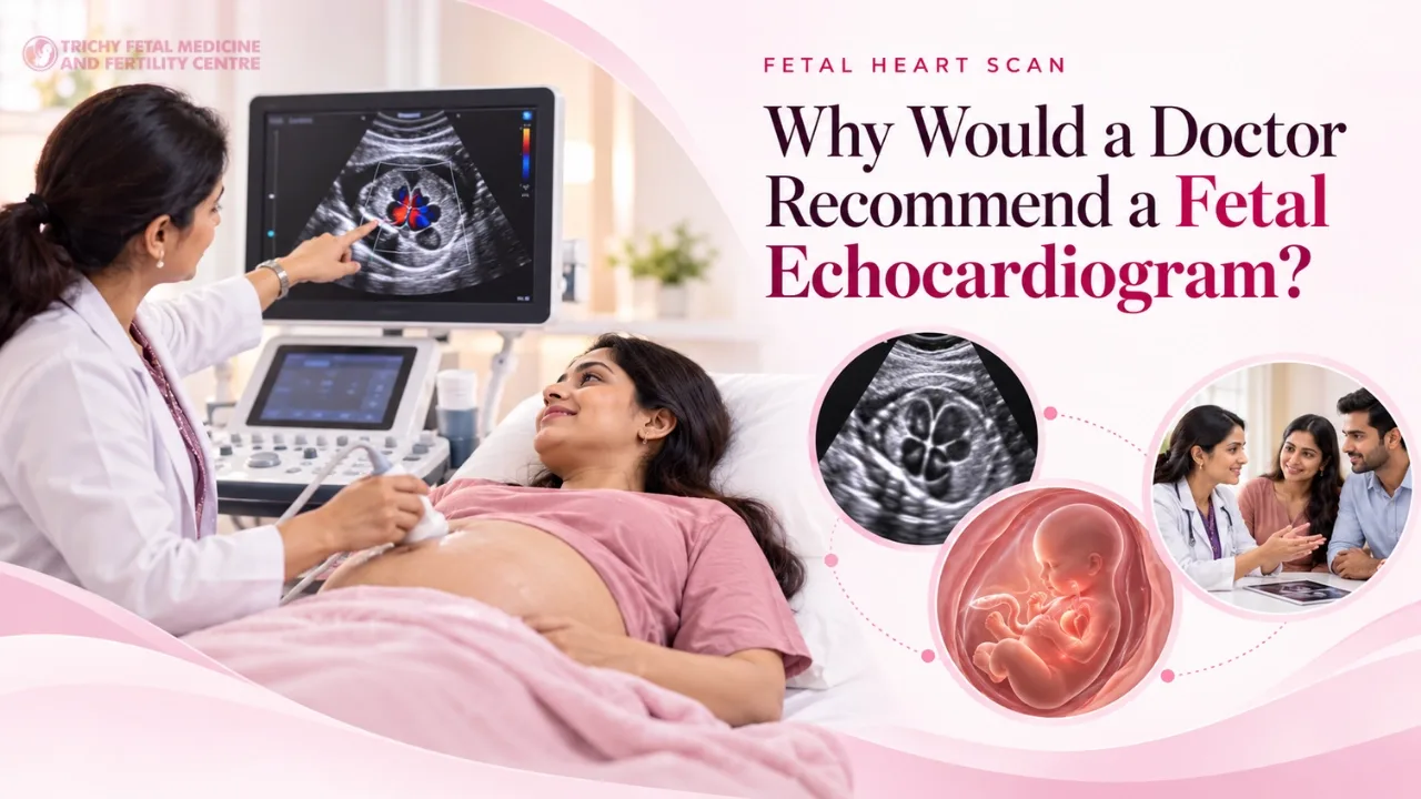
A fetal echo is a detailed heart scan of the unborn baby. Learn the common reasons doctors recommend it, including family history, diabetes, IVF and scan findings.
A fetal echocardiogram, or fetal echo, is a specialised ultrasound that examines the structure and function of the unborn baby's heart in detail. It goes beyond the heart views in a routine anomaly scan. Being referred for one can feel worrying, but most babies who have a fetal echo have normal hearts.
Why do heart defects matter?
Congenital heart defects are among the most common birth defects. Some are minor, while others need treatment soon after birth. Detecting them before birth allows parents to receive counselling and helps teams plan delivery at a centre with appropriate cardiac care.
What are the common reasons for referral?
Findings on routine scans
A concern seen in the heart views during the anomaly scan or NT scan is a frequent reason.
Increased nuchal translucency
An increased NT measurement at 11 to 14 weeks is associated with a higher chance of heart defects, even when chromosomes are normal.
Family history
A parent or previous child with a congenital heart defect increases the chance of a heart defect in the baby.
Maternal diabetes
Pre-existing diabetes, especially when poorly controlled in early pregnancy, raises the risk of heart defects.
IVF pregnancy
Pregnancies conceived through IVF or ICSI may have a slightly increased chance of heart defects in some studies.
Medicines and infections
Some medicines, such as certain anti-epileptic drugs, and infections such as rubella may affect heart development.
Abnormal heart rhythm
A very fast, slow or irregular fetal heart rate needs detailed assessment.
Monochorionic twins
Twins sharing a placenta have a higher chance of heart problems.
Chromosomal or genetic conditions
Known or suspected conditions linked with heart defects.
Maternal autoimmune conditions
Certain antibodies, such as anti-Ro, can affect the baby's heart rhythm.
When is a fetal echo done?
It is commonly done between about 18 and 24 weeks, when heart structures are easier to see. Some centres do an early heart assessment around 13 to 16 weeks in high-risk cases, followed by a later scan.
What should you expect?
The scan is similar to a regular ultrasound but takes longer and focuses on the heart's chambers, valves, vessels and rhythm. It is safe and non-invasive.
The short version
A referral for a fetal echo is a precaution that gives you clear information. Early detection supports planning and peace of mind. Dr Malathi G. Prasad offers fetal echocardiography and fetal medicine care in Trichy.
To arrange this at our Trichy centre, see fetal echo; if you are unsure whether you need it, our doctors can tell you after a review.