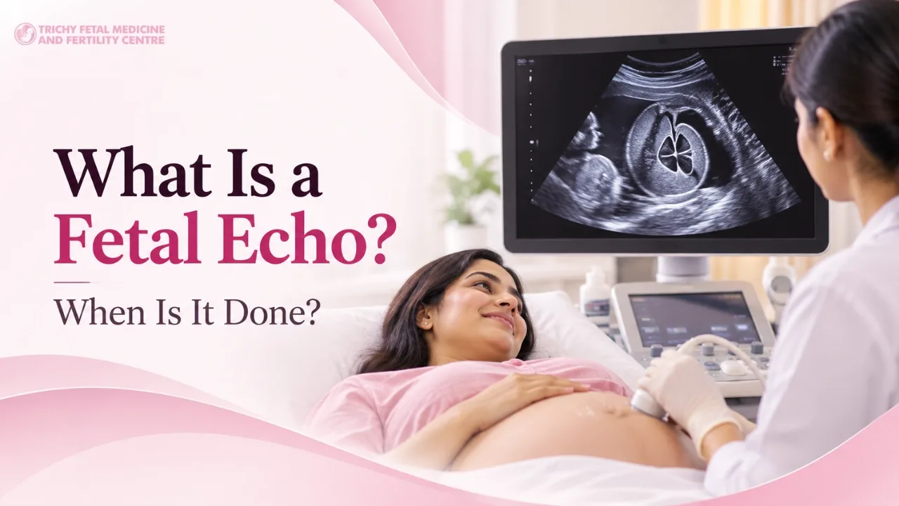
The routine anomaly scan looks at the whole baby, including a basic view of the heart. A fetal echo goes much further. It studies the heart chambers, valves, major vessels, blood flow and heart rhythm in detail.
When Is It Done?
The best window is usually 18 to 24 weeks, when the heart is large enough to be seen clearly. In some high-risk situations an early fetal echo may be done at about 12 to 16 weeks, and it can be repeated later if needed.
Who may need a fetal echo
A routine scan suggested a possible heart concern.
A previous baby, or a parent, has a congenital heart condition.
The mother has diabetes before pregnancy.
The mother has certain autoimmune conditions.
The pregnancy was conceived through assisted reproduction.
There is an abnormal NT scan or a chromosomal concern.
A twin pregnancy with shared placenta.
Exposure to certain medicines during pregnancy.
What happens during the test
The test is like a regular ultrasound. Gel is applied to the abdomen and a probe is moved over it. It is painless and safe for mother and baby. It can take 30 to 60 minutes, as the baby's position matters.
What do the results mean?
A normal result is reassuring. If a heart problem is found, your doctor will explain what it is, how it may affect the baby, where delivery should be planned and what treatment is likely after birth. Knowing early allows better planning.
What to do next
Most babies have a healthy heart, but a fetal echo gives answers when there is a reason to look closer. Dr Malathi G. Prasad at Trichy Fetal Medicine and Fertility Centre in Trichy can advise whether you need one and explain the findings clearly.
If this applies to your pregnancy, our fetal echo page explains what the visit involves, or you can meet our specialists before you decide anything.